Department of Neurosurgery
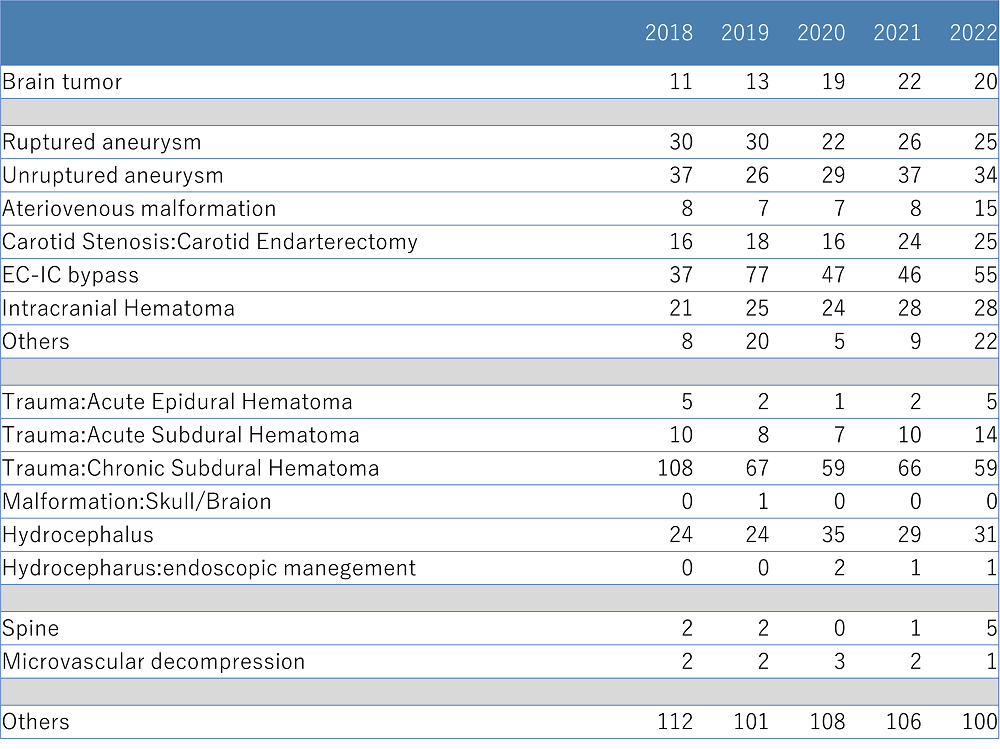
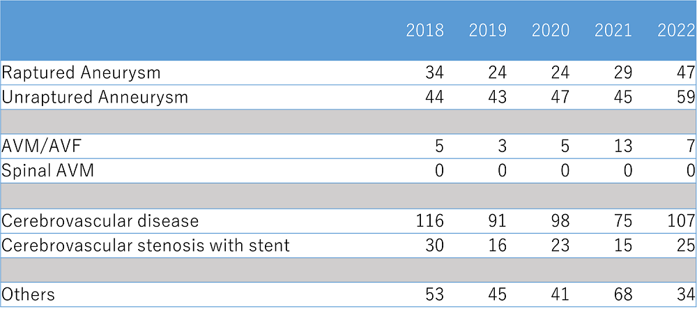
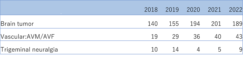
Target disease,treatment
Number of operations
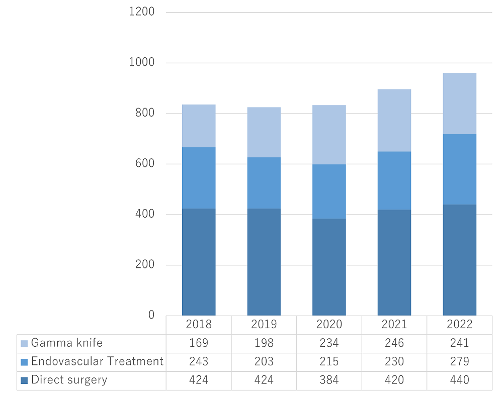
Surgical procedures
Endvascular treatment
gamma knife
Photographs and illustrated explanations of representative treatment techniques
Clipping of an unruptured anterior communicating artery aneurysm
Microscopic photographs of a surgical intervention for an unruptured middle cerebral artery aneurysm
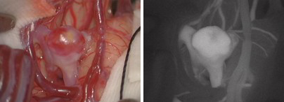
The image on the left shows a brain aneurysm before clipping. A tumor can be seen in a cerebral blood vessel.
The image on the right shows intravenous injection of a fluorescent pigment. The aneurysm and normal blood vessels appear white.
The image on the left shows clipping at the root (cervix) of the aneurysm.
The image on the right was captured after intravenous injection of a fluorescent pigment. Normal blood vessels appear white but not the aneurysm, indicating that blood flow to the aneurysm has been blocked.
Combination therapy for AVM
To treat AVM (a mass of blood vessels in the brain), for which surgical intervention is difficult, we often perform preoperative embolization (blocking blood vessels that are hard to treat surgically in advance) through endovascular surgery in combination with craniotomy or gamma knife radiosurgery, as in this case. Recently, ONYX®, a liquid material for embolization, has also become available.
- (1) Arteriovenous malformation (before operation): Malformation was found at two locations indicated with orange and white circles in the image. The malformation labeled with the orange circle was treated using a combination of endovascular treatment and craniotomy.
- (2a) After embolization: The malformation indicated with the orange circle considerably reduced in size.
- (2b) After embolization: ONYX® was injected in the blood vessels that appear as black lines within the orange circle.
- (3) Two days after embolization, the arteriovenous malformation was completely extracted through surgery. The malformation indicated with the white circle was treated with gamma knife radiosurgery at a later date, and follow-up observation is ongoing.
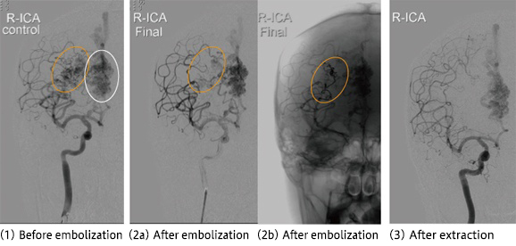
As illustrated in this case, we treat arteriovenous malformations with extraction via craniotomy, embolization, or gamma knife radiosurgery, or a combination of these techniques, for which we have adequate equipment and specialized physicians.
Treatment of Moyamoya disease
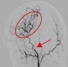
There are two types of Moyamoya disease: cerebral ischemia type and cerebral hemorrhage type. Bypass surgery is effective for the former type. At the NCVC, we actively perform bypass surgery to treat Moyamoya disease. The image on the left shows blood vessels in the brain after bypass surgery, demonstrating ample blood supply to the middle cerebral artery region (arrow) via the bypass (arrow).
Brain tumor
In addition to cerebrovascular diseases, the NCVC actively treats brain tumors, including meningiomas, schwannomas, and metastatic brain tumors.
The brain tumors that were recently treated are as follows (from left to right): parasagittal meningioma, angioblastoma, and vestibular schwannoma.
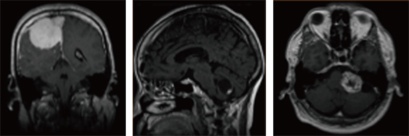
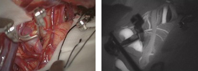
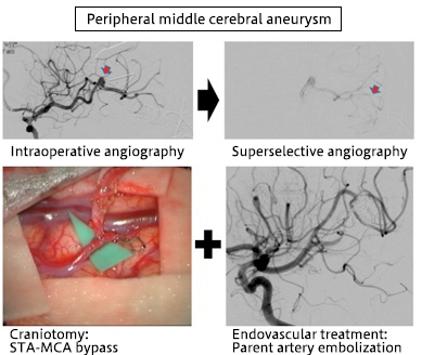
An example of hybrid surgery in a hybrid operating room
In January 2011, the NCVC commenced the use of a full-scale hybrid operating room, the first in Japan. A hybrid operating room is used for performing hybrid surgery, which involves a combination of craniotomy and endovascular treatment, two therapeutic methods with completely different concepts. Hybrid surgery has always been considered indispensable to treat hard-to-handle areas while ensuring safety and success. However, hybrid surgery is difficult to perform in conventional operating rooms that are separate from angiography rooms due to various limitations. A hybrid operating room combines the features of both the rooms, serving as a test for next-generation neurosurgery. At the NCVC, we operated on 15 patients in the hybrid operating room during its inaugural year. These comprised cases in which affected areas were hard to reach using the standard endovascular treatment, cases in which affected areas were difficult to identify using craniotomy, and highly invasive cases. The outcomes were favorable in all 15 cases. The hybrid operating room has also been used for the treatment of heart and aortic diseases. We believe that this operating room will be more actively used in the future not only for the treatment of hard-to-reach areas but also for achieving improved outcomes regarding safety and overall success.
Cerebral aneurysm coil embolization (endovascular treatment)
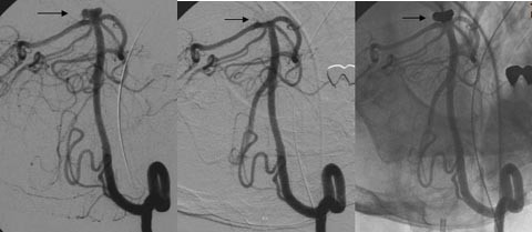
The angiographic image on the left shows an example of coil embolization of an unruptured cerebral aneurysm. Aneurysm (arrow) can be observed at the tip of the basilar artery. For examination, a microcatheter, which is thinner than a regular catheter, is carefully inserted into the aneurysm. A platinum coil is then introduced into the aneurysm via the microcatheter.
Images in the middle and on the right show the same region after coil embolization. There is no contrast medium inside the aneurysm.
Carotid artery stenting (CAS; an endovascular treatment)
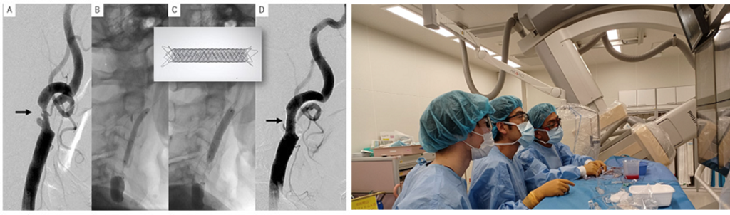
Images show a stent (metal tube) inserted to treat internal carotid artery stenosis at the cervix; note that the narrowed section (circled) has expanded.
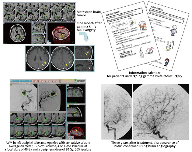
Gamma knife radiosurgery
The NCVC commenced gamma knife radiosurgery in April 2002; since then, many patients have been treated at the center with this modality. In February 2009, the equipment was upgraded to Model 4C, thereby largely reducing the time required for the procedure.
last updated : 2023/01/19